Can An Mr Chart Have A Negative Lcl - If the calculated control limit is farther from the center line than the value that you specify, minitab displays the bound. We know that defects cannot be less than zero (that is. You can start calculating the control limits after five data points. Lower control limit (lcl) the lcl is the greater of the following: The calculated average is then. The tool also seamlessly integrates. The control chart xmr consists of two charts: Another option is to use. At least for an individuals chart calculated this way. Individuals and moving range chart.

Control Chart Calculating Ucl And Lcl A Visual Reference of Charts
In minitab you can change the lower boundary to requested limit bound. 95% or 99% of data should fall within ucl and lcl. Another option.
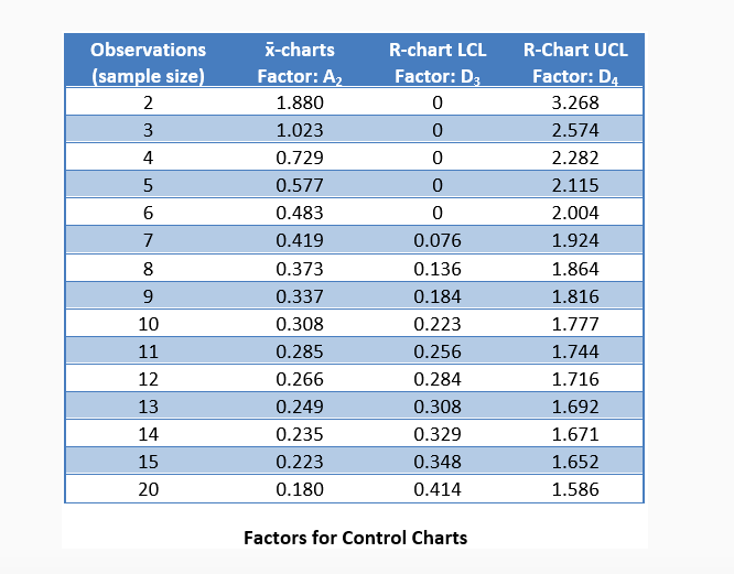
Solved Observations Kcharts Rchart LCL RChart UCL sample
Where mri is the moving. The control chart xmr consists of two charts: The calculated average is then. Determining whether a process is stable and.

Control charts for HPLC method used for the insulin quantification in
The average moving range, , of length w is given by the following formula: As we know sometimes when we calculate the natural process limits,.

IMR chart showing median waiting time of patients in the emergency
Draw the average line on the moving range chart. The lower control limit, labelled lcl on the graph, indicates that on this xbar chart, any.

Control Chart Calculating Ucl And Lcl A Visual Reference of Charts
This is then where you get a negative control limit. The median moving range is impacted much less by large moving range values than the.
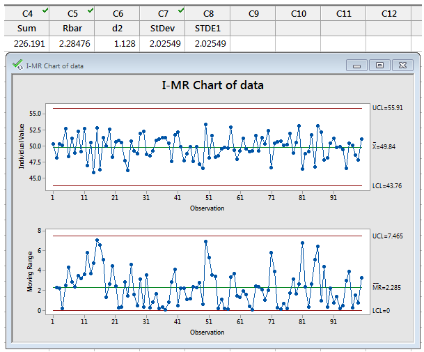
Methods and Formulas How Are IMR Chart Control Limits Calculated?
When you change an unstable process,. If the calculated control limit is farther from the center line than the value that you specify, minitab displays.

Imaging Characteristics of the Proximal Lateral Collateral Ligament of
You can start calculating the control limits after five data points. The control chart xmr consists of two charts: In some measures, that’s not a.
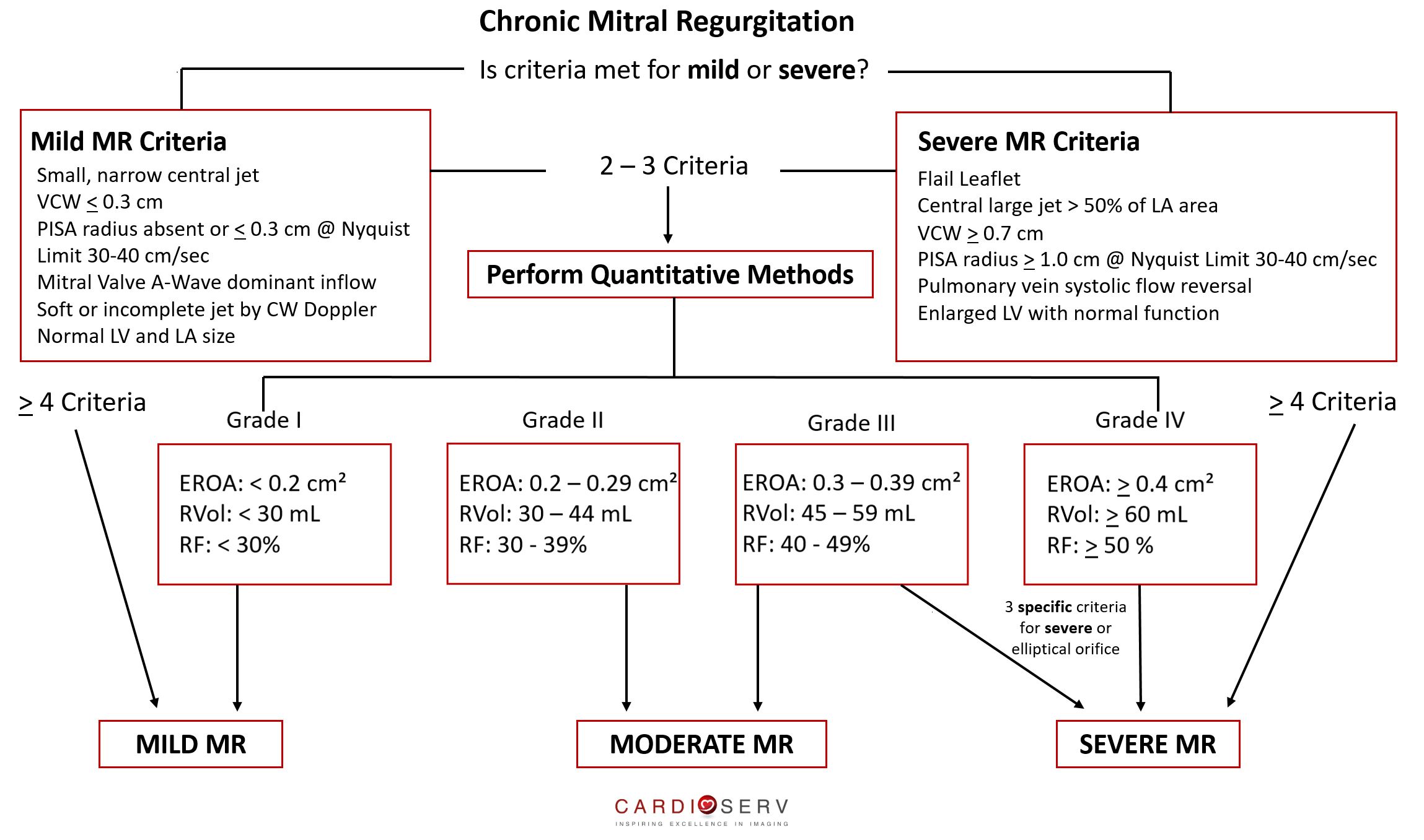
Mitral Regurgitation Case Study Demonstration! Cardioserv
Lower control limit (lcl) upper control limit (ucl) notation. Lower control limit (lcl) the lcl is the greater of the following: At least for an.

Lateral Collateral Ligament Complex
To calculate the upper control limit, multiply the average moving range, , by. Select the method or formula of your choice. 95% or 99% of.

Control Chart Calculating Ucl And Lcl A Visual Reference of Charts
Lower control limit (lcl) the lcl is the greater of the following: The control chart xmr consists of two charts: The lower control limit, labelled.
Upper Control Limit (Ucl) Notation.
Another option is to use. In minitab you can change the lower boundary to requested limit bound. If minitab plots the upper and lower control limits (ucl and lcl). Calculate the control limits for the moving range chart 1.
If The Lcl Comes Out Negative In Calculation, Then There Is No Lower Control Limit And Lcl Is Considered To Be Zero.
There are seven main types of control charts (c, p, u, np, individual moving range xmr, xbarr and xbars.) plus there are many more variations for special circumstances. Individuals and moving range chart. The average moving range, , of length w is given by. The median moving range is impacted much less by large moving range values than the average.
The Lower Control Limit, Labelled Lcl On The Graph, Indicates That On This Xbar Chart, Any Group Of Five Packages Averaging Under 41.7503G Is An Indication That The Process Is Unstable And Special Cause Variation Exists.
2 best practices when thinking about a lower control limit. To calculate the upper control limit, multiply the average moving range, , by. 95% or 99% of data should fall within ucl and lcl. Lower control limit (lcl) upper control limit (ucl) notation.
Select The Method Or Formula Of Your Choice.
You can specify a lower bound and an upper bound for the control limits. We know that defects cannot be less than zero (that is. In some measures, that’s not a practical value, like in the example below. Where mri is the moving.